CHEMICAL CODING
Most evolutionary scientists believe that the first sensory system of the earliest animal was a chemical sensitivity. Before the neocortex (Forebrain) evolved, there were areas of the midbrain and hindbrain devoted solely to sensing. Information on the Cranial Nerves are listed in a chart of the book on pg 92, table 4.4; The cranial nerves are illustrated on pg 85 (unit 5.7) of the coloring book.
What are the functions associated with the following cranial nerves (CN):
CN I.
CN II.; III, IV AND VI
CN V. AND VII
CN VIII
CN IX AND XII
CN X AND XI
STIMULUS
Organic substances containing chemicals that are perceived as sweet-sucrose, sour-HCL, bitter-quinine, salty-NaCl and umami or meaty-MSG. A combinaion of activity in five kinds of receptors (along with smell) give the perception of the taste of the food.
Taste buds are contained in papillae Taste receptors have excitable membranes and release neurotrnsmitters to excite other cells. Taste receptors are regenerated and replaced every 10-14 days, which is why food may taste more intense after an illness or after an extended fast. Taste receptors are located inside of tastebuds which are located in the papillae of the tongue.
TRANSDUCTION
When a substance tastes salty for instance, it means that a salitness receptor has detected the presence of sodium. Sodium ions cross into the membrane and produces an action potential. Sweetness, bitterness and umami receptors resemble the action of metabotropic receptors and therefore activate G-protein molecules and second messengers within the cell.
TRANSMISSION
Although each receptor detects
one kind of taste, several receptors create a particular firing pattern
that is perceived as a single taste experience. Information from the
receptors in the anterior 2/3 of the tongue synpase with the Chorda Tympani nerve and information from the posterior tongue and the throat travel along branches of the Cranial Nervs IX and X. What are the names of Cranial Nerves IX and X?
Taste nerves project to the nucleus of the solitary track (NTS) in the medulla. From the NTS, the information branches out to the pons, the lateral thalamus, the amygdala and the ventral posterior thalamus (VPN), finally terminating into areas of the cerebral cortex. The somatosensory cortex is where touch or food texture is detected on the tongue and the insula or primary taste cortex is where taste is perceived.. Innervation is ipsilateral in this sensory modality.
SENSATION, PERCEPTION AND COGNITION
The sensation of taste can be affected by culture and familiarity but also by genes and hormones. Give examples from the textbook on how genetic differences affect taste. Also how do hormones affect taste preferences, according to the text?
PUT IT ALL TOGETHER
For
the final exam, you should know the answer to the questions in red
above. Also you should be able to associate the following terms within
the correct cell of the class schema.
NaCl
Nucleus of the Solitary Tract
papillae
Primary gustatory cortex
saliva
taste buds
taste receptors
teeth
tongue
unami
Vagus Nerve
VEntral Posterior nucleus
This is the blog for the course Psyc 412- Physiological Psychology at Virginia State University
Tuesday, April 9, 2013
Thursday, April 4, 2013
Module 7.1 Audition
I. STIMULUS

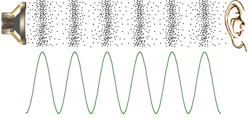
Sound waves are most often the result of periodic compressons of air. THe frequency of a sound is the number of compresssions per second, measured in Hz. Pitch is related to frequency such that higher pitch means higher frequency of the sound wave.
The amplitude of the sound wave related to its intensity and is related to how loudly the sound is perceived. Most adults hear sounds between 15,000-20,000Hz.
1. Identify the frequency and amplitude of the illustrated sound wave.
The compression of air is collected by the outer ear, turned into vibration in the middle ear and transduced in the inner ear.
2. Can you label the structures of the outer and middle ear.

II. TRANSDUCTION

Transduction happens in the cochlear, due to the movement of hair cells on the basilar membrane
.
III. TRANSMISSION PATHWAYS IN THE BRAIN

IV. SENSATION, PERCEPTION, COGNITION
Read and summarize this paper
http://www.nature.com/nature/journal/v416/n6876/full/416012a.html
Auditory System Site #1
Go to section 12.2 Sound: Intensity, Freuency, Outer and Middle Ear Mechanisms, Impedance Matching by Area and Lever Ratios.
You are only responsible for understanding the following terms for Section 12.2
*pinna,
*external auditory meatus (ear canal)
*middle ear ossicles - anvil, hammer, stirrup
*tympanic membrane
*oval window
round window
Auditory System site #2
Go to section 13.1- Figure 13.1 and 13.2 and hit "Play" Use this illustration (and your coloring book Figure 8.3). You are responsibile for the pathway structures and functions outlined in your coloring book.
USE THE MARKED TERMS ABOVE (*) AND THE TERMS BELOW IN THE HOMEWORK ASSIGNMENT- CLASS SCHEMA THAT IS DUE APRIL 3, 2014
*15-20,000 Hz
*superior olive
*medial geniculate of the thalamus
*inferior collicullus
*primary auditory cortex
*secondary auditory cortex
*pitch

Sound waves are most often the result of periodic compressons of air. THe frequency of a sound is the number of compresssions per second, measured in Hz. Pitch is related to frequency such that higher pitch means higher frequency of the sound wave.
The amplitude of the sound wave related to its intensity and is related to how loudly the sound is perceived. Most adults hear sounds between 15,000-20,000Hz.
1. Identify the frequency and amplitude of the illustrated sound wave.
The compression of air is collected by the outer ear, turned into vibration in the middle ear and transduced in the inner ear.
2. Can you label the structures of the outer and middle ear.

II. TRANSDUCTION

Transduction happens in the cochlear, due to the movement of hair cells on the basilar membrane
.

III. TRANSMISSION PATHWAYS IN THE BRAIN

IV. SENSATION, PERCEPTION, COGNITION
Read and summarize this paper
http://www.nature.com/nature/journal/v416/n6876/full/416012a.html
V. PUT IT ALL TOGETHER- Stimulus, Transduction, Transmission, Sensation, Perception & Cognition
If you are really understand this material, you should be able to easily read the following webites. Once you are comfortable reading the websites below, your next assignment will be to complete the Audition portion of the Sensory Modality chart.
Auditory System Site #1
Go to section 12.2 Sound: Intensity, Freuency, Outer and Middle Ear Mechanisms, Impedance Matching by Area and Lever Ratios.
You are only responsible for understanding the following terms for Section 12.2
*pinna,
*external auditory meatus (ear canal)
*middle ear ossicles - anvil, hammer, stirrup
*tympanic membrane
*oval window
round window
Auditory System site #2
Go to section 13.1- Figure 13.1 and 13.2 and hit "Play" Use this illustration (and your coloring book Figure 8.3). You are responsibile for the pathway structures and functions outlined in your coloring book.
USE THE MARKED TERMS ABOVE (*) AND THE TERMS BELOW IN THE HOMEWORK ASSIGNMENT- CLASS SCHEMA THAT IS DUE APRIL 3, 2014
*15-20,000 Hz
*superior olive
*medial geniculate of the thalamus
*inferior collicullus
*primary auditory cortex
*secondary auditory cortex
*pitch
Monday, April 1, 2013
Module 6.1, 6.2 & 6.3- Visual System Anatomy
I. THE STIMULUS
A. Getting the image to the retina
Most of the structures of the eyeball are involved in preparing the image that is reflecting visible light into the eye. The visible light range of the electromagnetic spectrum is the frequency of approximately 400-700nm. Humans perceive the shortest visible wavelengths as violet, medium short wavelenths is green; medium long wavelength is perceived as yellow and long wavelenght perceived as red. (see Figure 6.8 on page 160). Once the particular range of the electromagnetic energy is reflected off the image, the pattern of the reflected image enters the eyeball.
1. What role do the iris, pupil, lens and cornea (structures of the eyeball) play in getting the pattern of the reflection onto the photoreceptors of the retina? (pag 156)
2. In the illustration below draw in the placement of horizontal cells and amacrine cells.(page 169)
3. Which cell axons leave the eyeball and what is this collection of axons called. Also, why is there a blindspot?
FRONT OF EYE BALL BACK of EYE BALL
4. What is the role of the fovea? (pg 157)
5. What is the functional significance of a midget ganglion cell?
6. What is convergence?
II. TRANSDUCTION (Retinal Processing)
IIIa. TRANSMISSION PATHWAYS IN THE EYE
Ganglion Receptive Fields (see lecture notes & pg 172)
IIIb. TRANSMISSION PATHWAYS IN THE BRAIN
Transmission of visual information actually begins in the retinal layers once the photoreceptors are stimulated.
1. Trace the transmission pathway from when retinal cell axons leave the eyeball to the destination synapse of MOST of those cells in the LGN .(see pg 168 textbook and pg 134 Coloring Book)
Lateral Geniculate Nucleus is located in the Thalamus. (See PAGE 98 of Coloring Book)
IV. SENSATION, PERCEPTION and COGNITION
a. Receptive Fields (Sensation and Perception)
Use this site to help your understand the concept of Receptive Fields. You should understand the sections labeled
THE RETINA
RECEPTIVE FIELDS FROM THE RETINA TO THE CORTEX
You are not responsible for the third section- THE CELLULAR STRUCTURE OF THE VISUAL CORTEX.
b. Cogntion
Shape,
Color Perception
Motion Perception
The following terms from this link will help you "put it all together" in the story of the sensation of Vision. USE THE FOLLOWING TERMS MARKED (*) FOR YOUR HOMEWORK ASSIGNMENT- VISION AND THE CLASS SCHEMA.
THE EYE
retina
*cornea
*lens
*photoreceptors
rods
cones
*amacrine
horizontal
*ganglion cells
*pupil
iris
first visual system synapse
THE TARGETS OF THE OPTIC NERVE
optic disk,
optic nerve
ganglion cell axons
*optic chiasm
*lateral geniculate nucleus of the thalamus
LGN receptive fields
THE VARIOUS VISUAL CORTICES
receptive fields of the cells of the retina
occipital lobe
*primary visual cortex
secondary visual cortex
*posterior inferior temporal cortex
*middle temporal cortex
*medial superior temporal cortex
ventral pathway
dorsal pathway
A. Getting the image to the retina
Most of the structures of the eyeball are involved in preparing the image that is reflecting visible light into the eye. The visible light range of the electromagnetic spectrum is the frequency of approximately 400-700nm. Humans perceive the shortest visible wavelengths as violet, medium short wavelenths is green; medium long wavelength is perceived as yellow and long wavelenght perceived as red. (see Figure 6.8 on page 160). Once the particular range of the electromagnetic energy is reflected off the image, the pattern of the reflected image enters the eyeball.
1. What role do the iris, pupil, lens and cornea (structures of the eyeball) play in getting the pattern of the reflection onto the photoreceptors of the retina? (pag 156)
2. In the illustration below draw in the placement of horizontal cells and amacrine cells.(page 169)
3. Which cell axons leave the eyeball and what is this collection of axons called. Also, why is there a blindspot?
FRONT OF EYE BALL BACK of EYE BALL
4. What is the role of the fovea? (pg 157)
5. What is the functional significance of a midget ganglion cell?
6. What is convergence?
II. TRANSDUCTION (Retinal Processing)
IIIa. TRANSMISSION PATHWAYS IN THE EYE
Ganglion Receptive Fields (see lecture notes & pg 172)
IIIb. TRANSMISSION PATHWAYS IN THE BRAIN
Transmission of visual information actually begins in the retinal layers once the photoreceptors are stimulated.
1. Trace the transmission pathway from when retinal cell axons leave the eyeball to the destination synapse of MOST of those cells in the LGN .(see pg 168 textbook and pg 134 Coloring Book)
Lateral Geniculate Nucleus is located in the Thalamus. (See PAGE 98 of Coloring Book)
IV. SENSATION, PERCEPTION and COGNITION
a. Receptive Fields (Sensation and Perception)
Use this site to help your understand the concept of Receptive Fields. You should understand the sections labeled
THE RETINA
RECEPTIVE FIELDS FROM THE RETINA TO THE CORTEX
You are not responsible for the third section- THE CELLULAR STRUCTURE OF THE VISUAL CORTEX.
b. Cogntion
Shape,
Color Perception
Motion Perception
V. PUT IT ALL TOGETHER- Stimulus, Transduction, Transmission, Sensation, Perception & Cognition
If you are really understand this material, you should be able to easily read the following webites. Once you are comfortable reading the websites below, your next assignment will be to upload your version of the "Sensory Stories," in which you will be able to explain this sensory modality from start to finish on a YouTube video. More about the Sensory Stories assignment in class.
Put it all together site for VisionThe following terms from this link will help you "put it all together" in the story of the sensation of Vision. USE THE FOLLOWING TERMS MARKED (*) FOR YOUR HOMEWORK ASSIGNMENT- VISION AND THE CLASS SCHEMA.
THE EYE
retina
*cornea
*lens
*photoreceptors
rods
cones
*amacrine
horizontal
*ganglion cells
*pupil
iris
first visual system synapse
THE TARGETS OF THE OPTIC NERVE
optic disk,
optic nerve
ganglion cell axons
*optic chiasm
*lateral geniculate nucleus of the thalamus
LGN receptive fields
THE VARIOUS VISUAL CORTICES
receptive fields of the cells of the retina
occipital lobe
*primary visual cortex
secondary visual cortex
*posterior inferior temporal cortex
*middle temporal cortex
*medial superior temporal cortex
ventral pathway
dorsal pathway
Subscribe to:
Posts (Atom)


