Tuesday, March 31, 2015

Textbook Module 7.2 Mechanical Senses- Somatosensation

Somatosensory Systems
The somatosensory system monitors the sensations of the body and its movements and include multiple systems.

STIMULUS
The different types of mechanical stimuli are listed in the text in Table 7.1 (pg 205). The stimuli include pain (generated by cell injury); heat or cold (on either side of physiological cell injury range)
Movement of hairs on the skin surface; sudden displacement of skin; light touch; skin stretch or also stretch or injury to joints and muscles.

 What these all have in common is that they are associated with types of receptors that are sensitive to them.

TRANSDUCTION
Receptors associated with the types of stimuli listed above are also listed in Table 7.1 They are
Free nerve endings; hair follicle receptors Meissner's corpuscles, Pacinian corpuscles, Merkel's disks, Ruffini endings and Krause end bulbs.  These receptors are located in the  skin and in the joints and muscles.  Stimulation of a touch receptor opens sodium channels in the axon and thereby starts an actionl potential.

TRANSMISSION
Somtosensory information from touch receptors on the head enter the CNS through the cranial nerves, such as Cranial Nerve V. . Somatosensory information  from receptors below the head enter the spinal cord via the dermatones.   Dermatomes were discussed previously in Module 2.1- cells of the Nervous System on this blog.

Transmission Pathway for Fine Touch and Vibratory Sense-  Dorsal Column Medial Lemniscus
see unit 9.5-9.6 of the Coloring Book (pg 142-145)


  • The transmission pathway for touch is carried into the spinal cord via the dorsal roots (see Unit 1.4 Coloring Book pg 10) and then without synapsing ascend ipsilaterally in the dorsal part of the spinal cord to the dorsal column nuclei.  Sensory input from the legs synapse in the nucleus gracilis and input from the arms synapse in the nucleus cuneatus. 
  • Axons from the dorsal column nuclei decussate in the medulla and ascend to the the thalamus via the dorsal column medial lemniscus.
  • Axons from the dorsal column medial lemniscus ascend to the thalamus.  Fibers from Cranial Nerve 5 join the medial lemniscus enroute to the thalamus
  • Most of the axons of the medial lemniscus synapse on neurons in the ventral posterior nucleus of the thalamus.
  • Axons from the VPN project to the primary somatosensory cortex (Coloring book unit 7.6 pg 120).
  • The primary somatosensory cortex is located on the post central gyri (see coloring book unit 7.1 pg 119) of the parietal lobes (CB unit 7.2 pg 112).
  • The output of the primary somtosensory cortex is mostly projected to the secondary somatosensory cortex located just inferior to the primary somatosensory cortex located in the ventral portion of the postcentral gyrus and hidden by the lateral fissure
  • Output from the sencodary somatosensory cortex is sent to the posterior parietal association cortex.


The somatosensory cortex is laid out somatopically, a map of the body is on the brain. (Just like there is a retinotopic representation of the visual image represented on the occiptial lobe and a tonatopic representation of frequency represented in the cochlea and auditory cortex).
The somatosnesory body map is called a homonculus. (CB unit 8.6 pg 144).


Transmission Pathway for Pain, Temperature, Crude Touch -Spinothalamic Tracts
Pain and Temperature (lateral spinothalamic tracts) Crude Touch (anterior spinothalamic tract)
 see Textbook figure 7.15 and 7.16 (pg 208-209).

Why is pain information conveyed so slowly to the brain?
What are the neurotransmitters involved in conveying pain in the spinal cord?
How does the pain transmission pathway differ from the touch transmission pathway?

Pain information crosses to the contralateral side of the spinal cord at once. The pathways then continue to the brain via the spinothalamic tract. This axonal pathway begins in spine then ends in thalamus- thus the name spine -o- thalamic.


  • The spinothalamic tract conveys pain and temperature (lateral spinothalamic tract) and crude touch (anterior spinothalamic tract).
  • Primary sensory nerves that register pain have  cell bodies that lie in the posterior dorsal root ganglion. 
  • They synapse in in the posterior horn of the dorsal root in the spinal cord.
  • Secondary neurons cross the spinal cord contralaterally and ascend to the brain via the  lateral spinothalamic tract in a somatotopic arrangement. 
  • Secondary neurons synapse in the VPN of the thalamus,
  • From the VPN of the thalamus, tertiary neurons ascend via the internal capsule to the primary sensory cortex  
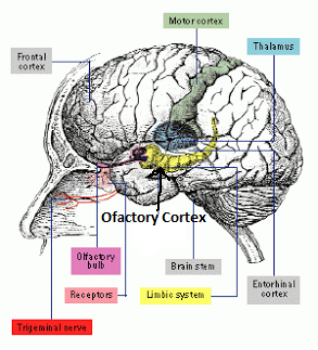
  • These cells also branch off axon collaterals to the limbic system, hypothalamus, and other portions of the CNS such s the amygdala, hippocampus and prefrontal cortex.  These areas react not to the sensation of pain but  to its emotional associations.

SENSATION PERCEPTION COGNITION

Descending Analgesic Circuit
Textbook Figure 7.18 pg 210 and unit 8.7 of the Coloring Book (pg 146)

A centrifugal pathway is a pathway that exerts either a facilliatory or inhibitory influence on a nerve signal.  One example of a centrifugal pathway is the descending analgesia circuit.  During times of intense emotion it is possible to feel little or no pain, even when there is a grave injury.

What is a centrifugal pathway?

The inhibition of pain information in the brain is due to the activation of natural opioids or endorphins in the midbrain structure, the periaqueductal gray (PAG)- gray area surrounding the cerebral aqueduct in the midbrain.

Axons from the PAG synapse back to the spinal cord  and decreases pain signals in the spinal cord. Therefore, even though pain receptors may be firing the signal to the brain has been blocked and therefore the injured individual does not experience pain.

See figure 7.18 in the textbook
  • "Certain kinds of painful and other stimuli" send emotional pain information from the cortex to the PAG via endorphine neurotransmitters.  
  • The PAG projects to the "area in the rostral part of the medulla" and excites cells that release more endorphine neurotransmitter into the spinal cord. 
  • The endorphine neurotransmitter inhibits the primary sensory nerve which is releasing substance P as a neurotransmitter and therefore stops "areas of the spinal cord that receive pain messages" from sending ascending pain messages to the brain
  • 'The injured person experiences no pain

More recent research about this pathway can be found here.


Tuesday, March 24, 2015

Textbook Module 7.1 Audition

I. STIMULUS

 http://www.mediacollege.com/audio/images/loudspeaker-waveform.gif
 Sound waves are most often the result of periodic compressons of air. THe frequency of a sound is the number of compresssions per second, measured in Hz. Pitch is related to frequency such that higher pitch means higher frequency of the sound wave.
The amplitude of the sound wave related to its intensity and is related to how loudly the sound is perceived. Most adults hear sounds between 15,000-20,000Hz.
1. Identify from the wave illustration which characteristic illustrates frequency and which characteristic illustrates amplitude.


The compression of air is collected by the outer ear, turned into vibration in the middle ear and transduced in the inner ear.
2. Can you label the structures of the outer and middle ear.


II. TRANSDUCTION


Transduction happens in the inner ear or cochlear. The chochlear is a fluid filled chamber deep in the ear.  When a pressure wave is created by movement of the oval window the basilar membrane moves and the hair cells located between the basilar and tectorial membranes also mimic the exact wave.

.


III. TRANSMISSION PATHWAYS IN THE BRAIN


IV. SENSATION, PERCEPTION, COGNITION

Read and summarize this paper for EXTRA CREDIT (10 points for a 1 page double spaced document in which the most important points of this paper are summarized.)  This assignment is due April 2, 2015.

http://www.nature.com/nature/journal/v416/n6876/full/416012a.html


V. PUT IT ALL TOGETHER- Stimulus, Transduction, Transmission, Sensation, Perception & Cognition

Know where the following terms fit on the class schema.


*pinna,
*external auditory meatus (ear canal)
*middle ear ossicles - anvil, hammer, stirrup
*tympanic membrane
*oval window
round window 
*15-20,000 Hz
*superior olive
*medial geniculate of the thalamus
*inferior collicullus
*primary auditory cortex
*secondary auditory cortex
*pitch


Monday, March 23, 2015

Textbook Module 7.3 Olfaction -Chemical Senses

STIMULUS
Olfaction is the response to chemicals that contact the membranes inside the nose. Olfaction is important in food selection, as most of what we call 'flavor" is a combination of the taste, texture and aroma of the food. The stimulus for olfaction are air-borne molecules that are ordorant chemicals

TRANSDUCTION
Olfactory cells line the nasal cavity. Each ell has cilia that extend from the cell body into the nasal passage. Olfactory receptors are located on the cilia.  Ordorant chemicals activate synaptic transmission. Olfactory receptors have synapses with cells in the olfactory bulb.

While there are only 3 types of cone cells that produce color vision and 5 types of taste receptors that provide flavor, there are several hundred types of olfactory receptors that respond to different types of chemicals. Olfactory receptors are vulnerable to damage since they are exposed to the air. The lifetime for such cells is just around a month.


TRANSMISSION
 When an olfactory receptor is stimulated the axon synapses in the olfactory bulb. Coding for the smell begins in the olfactory bulb. What does the book say about the changes in olfactory bulb cell firing based on whether the scent is different or more intense?  KNOW FOR FINAL EXAM

Cells of the olfactory bulb axons synapse in the olfactory area of the cerebral cortex known as the olfactory cortex.


SENSATION PERCEPTION COGNITION
Olfaction also plays a role in social behavior.  What are the scent-related chemicals involved in social behavior?  Who detects orders more readily? Men or Women -- KNOW FOR FINAL EXAM

SYNESTHESIA
     What is synesthesia?  What sensory systems are identified in the textbook explaination of synesthesia?  KNOW FOR FINAL EXAM


 PUT IT ALL TOGETHER
For the final exam, you should know the answer to the questions in red above.  Also you should be able to associate the following terms within the correct cell of the class schema.

air borne chemical ordorants
medial dorsal nucleus of the thalamus
nasal passages 
Olfactory bulbs
olfacotry glomeruli
Olfactory protein molecule
olfactory receptor cells
orbitofrontal cortex
a rose

Sunday, March 22, 2015

Textbook Module 7.3 Gustation- Chemical Senses

CHEMICAL CODING
Most evolutionary scientists believe that the first sensory system of the earliest animal was a chemical sensitivity.  Before the neocortex (Forebrain) evolved, there were areas of the midbrain and hindbrain devoted solely to sensing.  Information on the Cranial Nerves are presented on page 84 (unit 5.7) of the coloring book.

What are the behavioral functions associated with the following cranial nerves (CN):
CN I.
CN II.;
CN V.
CN VIII

KNOW THE ANSWER TO THIS QUESTION FOR FINAL EXAM

STIMULUS
 Organic substances containing chemicals that are perceived as sweet-sucrose, sour-HCL, bitter-quinine, salty-NaCl and umami or meaty-MSG.  A combination of activity in five kinds of receptors (along with smell) give the perception of the taste of the food.

Taste buds are contained in papillae Taste receptors have excitable membranes and release neurotrnsmitters to excite other cells. Taste receptors are regenerated and replaced every 10-14 days, which is why food may taste more intense after an illness or after an extended fast.  Taste receptors are located inside of tastebuds which are located in the papillae of the tongue.

TRANSDUCTION
When a substance tastes salty for instance, it means that a salitness receptor has detected the presence of sodium. Sodium ions cross into the membrane and produces an action potential.  Sweetness, bitterness and umami receptors resemble the action of metabotropic receptors and therefore activate G-protein molecules and second messengers within the cell.


TRANSMISSION
Although each receptor detects one kind of taste, several receptors create a particular firing pattern that is perceived as a single taste experience. Information from the receptors in the anterior 2/3 of the tongue synpases with the Chorda Tympani nerve and information from the posterior tongue and the throat travel along branches of the Cranial Nervs IX and X.  What is the name of Cranial Nerve X ? 


 Taste nerves project to the nucleus of the solitary track (NTS) in the medulla. From the NTS, the information branches out to the pons, the lateral thalamus, the amygdala and the ventral posterior thalamus (VPN), finally terminating into areas of the cerebral cortex. The somatosensory cortex is where touch or food texture is detected on the tongue and the insula or primary taste cortex is where taste is perceived..  Innervation is ipsilateral in this sensory modality. 


SENSATION, PERCEPTION AND COGNITION
  The sensation of taste can be affected by culture and familiarity but also by genes and hormones. Give examples from the textbook on how genetic differences affect taste. Also how do hormones affect taste preferences, according to the text?  This is an extra credit opportunity and is due on April 2, 2015


 PUT IT ALL TOGETHER
 You should be able to associate the following terms within the correct cell of the class schema.

 NaCl
Nucleus of the Solitary Tract
papillae 
Primary gustatory cortex
saliva
taste buds
taste receptors
teeth
tongue 
unami
Vagus Nerve
Ventral Posterior nucleus