
Tuesday, October 13, 2015
Friday, September 4, 2015
Textbook Module 2.1 Coloring book assigments and Vocabulary Terms Complete List
Textbook Module 2.1 The concept of the Synapse
The important thing about this chapter is the idea of a synaptic network of 3 neurons. We identified them as Cell A, B and C. You should have indicated this network in the coloring book from the exercise done in class.
The first actual network described in class was the reflex arc.
For this text book module:
Please connect the book figures and these coloring book pages./ Taken from Blackboard
BY THE MIDTERM EXAM YOU SHOULD KNOW THE FOLLOWING TERMS AND THEIR DESCRIPTION AND LOCATION (* =terms used by the professor). REMEMBER THAT IN THIS CLASS WE ARE VIEWING THE SAME TOPICS THROUGH DIFFERENT LEVELS OF ANALYSIS. (i.e. BIG PICTURE,-nervous system anatomy SMALL PICTURE individual nerurons, BIGGEST PICTURE behavior AND TEENY-TINY PICTURE synaptic transmission
The important thing about this chapter is the idea of a synaptic network of 3 neurons. We identified them as Cell A, B and C. You should have indicated this network in the coloring book from the exercise done in class.
The first actual network described in class was the reflex arc.
For this text book module:
Please connect the book figures and these coloring book pages./ Taken from Blackboard
| Neurons and other Cells of the Nervous System | TEXTBOOK | COLORING BK |
| Neural morphology | Fig 2.5 pg 31 | CB 3.1 pg 32 |
| Cell organelles | Fig 2.2 pg 29 | CB 3.2-3.3 pg 34-36 |
| Lipid bilayer and receptors | Fig 2.3 pg 30 | CB 3.7 pg 44 |
| Myelination | CB 3.6 pg 42 | |
| Glia Cells | Fig 2.10 pg 33 | Fig 3.8 pg 46 |
BY THE MIDTERM EXAM YOU SHOULD KNOW THE FOLLOWING TERMS AND THEIR DESCRIPTION AND LOCATION (* =terms used by the professor). REMEMBER THAT IN THIS CLASS WE ARE VIEWING THE SAME TOPICS THROUGH DIFFERENT LEVELS OF ANALYSIS. (i.e. BIG PICTURE,-nervous system anatomy SMALL PICTURE individual nerurons, BIGGEST PICTURE behavior AND TEENY-TINY PICTURE synaptic transmission
| Astrocytes | Axon | Axon hillock |
| Buttons, Terminal buttons* or axon boutons* | Cell body or *Soma | Cell membrane or *Lipid Bilayer |
| Cytoplasm | Dendrites | Dendritic spines |
| Dendritic arborization* | G-protein | Golgi apparatus |
| Interstitial Fluid | Ionotropic receptors | Macroglia |
| Ion channels | Microglia | Microtubules |
| Metabotropic receptors | Neurofilaments | Neurotransmitter molecule |
| Nodes of Ranvier | Nucleus | Oligodendrocytes |
| Post-synaptic membrane | Presynaptic membrane | Receptive area |
| Receptors | Ribosome | Endoplasmic reticulum |
| Schwann cells | Signal proteins | Synapse |
| Synaptic vesicles |
Tuesday, August 25, 2015
Neuroanatomy, Neurophysiology and Neuropharmacology: The Basics
I. Brief, Basic Chemistry Review (Appendix A- pg 486)
A. Elements and Compounds
1. As stated in Chapter 1, the Big Bang initially produced 4 forces- 1. gravity, 2. electromagnitism 3. strong nuclear forces and 4. weak nuclear forces. Chemistry is the study of the weak nuclear forces found in compounds. The weak nuclear forces allow for chemical reactions which are the transfer of ions and molecules to make new molecules and compounds. When wood is burned it turns into ashes and gases and water vapor and also produces heat. These are all chemical reactions. The basis of chemical reactions is the interaction of elemental chemicals. The periodic table lists 92 elements of which only are few are found in living organisms. Some of the most important are listed here.
Q. What are the most important elements in living organisms?
2. Compounds are made from molecules which are made from elements. Elements that are listed on the Periodic Table are comprised of atoms. For instance, when listed on the Periodic Table, Hydrogen has an atomic number of #1 and Oxygen is #16. The atomic number corresponds to the number of protons in the nucleus of an atom of that element. It also corresponds to the number of electrons in the neutral atom. When atoms come together, they make molecular compounds. Water is a compound or molecule. Compounds are created when electrons from different atoms join each other in chemical reactions. For instance, the molecules of water are elements Hydrogen and Oxygen. On each water molecule are two atoms of hydrogen and 1 atom of oxygen (H20). Each atom is composed of subatomic particles, protons, neutrons and electrons, which are positively charged, neutral and negatively charged- in that order. Protons and neutrons are found in the center of the atom, the nucleus and electrons are found in the space around the nucleus. It is very very difficult to separate protons which is a property of strong nuclear forces. The result of breaking those nuclear bonds is an atomic bomb. In a stable state, an atom will have the same number of protons as electrons. Molecules prefer a stable state to an unstable state and this is what drives ma
Q. What is the atomic number for Oxygen, Hydrogen and Carbon? What do these numbers mean?
B. Ions
3. An atom that has gained or lost one or more electron is called an ion. When the compound salt NaCl (Sodium and Chloride) is placed in water it dissolves or dissociates into ions: Na+ and Cl-. Sodium atoms lose electrons and become positively charged (has space available to accept an electron). A negatively charged ion, like Cl- has an extra electron. Thus ions with opposite charges attract each other and ions with the same charge repel each other. This is a property of electromagnitism and contributes to the property of weak nuclear forces.
In some cultures physics and chemistry are taught before biology because these fields underlie the very complex mechanisms necessary for biological systems. The topics above provide an extremely brief background for processes that are involved in synaptic transmission.
Friday, August 21, 2015
Textbook Module 1.2 Genetics and Behavior
Module 1.2 Genetics and Behavior January 20, 2015
PLEASE READ THE ENTIRE MODULE 1.2 FROM THE TEXTBOOK AND THEN READ THESE NOTES. PAY PARTICULAR ATTENTION TO THE MATERIAL IN RED THAT IS BELOW. THE RED TEXT MAY BE USED FOR QUIZ/TEST QUESTIONS.Mendelian Genetics
The book concludes that it is both Nature and Nurture since the Module begins with the statement "Everything you do depends on both your genes and your environment."
The evidence provided for this statement is facial expressions. What kind of observations led to the conclusion about facial expressions? Other behaviors listed are intelligence, sexual orientation, alcoholism and weight gain. Could you provide evidence for both genetic and environmental influences on these behaviors?
I understand that many of you may have taken Biology some time ago, so you may be unfamiliar with Mendelian Genetics. I've tried to provide a brief but effective re-introduction, starting with this video.
A much broader view of Figure 1.7 and Figure 1.9
Here you will be reminded that living tissue is made up of cells. Every cell has a nucleus, Every nucleus contains materials for the replication of the cell, genes. Genes are made up of DNA. RNA is used to replicate the gene from the DNA.
According to the text, Genes are units of heredity that maintain their structural identity from one generation to another. Genes are defined as a portion of a chromosome which is composed of the double-stranded molecule DNA. Here's a movie to help visualize this. (Please watch the videos SEVERAL times in order to answer the questions in red below.)
Genes are reproduced through a process of DNA replication and DNA transcription. A strand of DNA serves as a template for the synthesis of RNA, a process called transcription.
From the Videos above answer the following questions.
1. could you explain how the following terms differ from one another?
DNA, Chromatin, Chromasomes, Histones, Nucleosome
2. How many molecules of DNA are synthesized by RNA.
Once there is a single strand of messageRNA or mRNA (copied DNA), the strand leaves the nucleus and is joined to ribosomes where amino acids sequences produce proteins. This process is translation.
Genes can be dominant, recessive or intermediate. Here's an explanation using what Mendel used, peas.
From the Video define- allele, genotype and phenotype.
The list of traits given at the beginning of the chapter include intelligence, sexual orientation, alcoholism and weight gain. Now that you have a clearer understanding of genotype and phenotype, do you believe there is a genetic basis for intelligence, sexual orientation alcoholism or weight gain. Be able to explain your answer?
Genetic Changes
Genes can change and therefore the transcription by mRNA will be altered as well as translation of proteins as the final outcome.
Mutations of the gene (incl. duplication and deletion)
Epigenetics- Various experiences can turn a gene on or off. Evidence of epigenetic effects come mainly from animal studies, several of which were described on pg. 12.
What did Godfrey et al, 2007 discover about obesity and heart disease?
What did Weaver et al, (2004) find out about the effects of maternal care on stress levels of rat pups?
How does an experience modify gene expression?
Heredity and Environment
Evidence about the influence of heredity and environment often come from twin studies. Why?
What does it mean to be a monozygotic or dizygotic twin?
Evolutionary Psychology
Define the terms: altruism, kin selection and reciprocal altrusim
Textbook Module 5.1 Neural Development
Neurogenesis
Please review the links to help understand the process.
ONLINE VIDEOS to Visualize Neural Tube
Coloring Book Assignment for Neurogenesis (Understand these processes)
CB 4.2 Processes of Early Neural Development
Stage 1- Proliferation
Stage 2- Migration
Radial Glial cells
Stage 3-Aggregation
Growth Cones and Circuit formation
Stage 4- Process Growth and Synapse Formation
Circuit pruning
Stage 5- Neuron Death
Stage 6 Myelination
CB 4.5 Axon Growth: Correctly Wiring the Nervous System
A. Chemoaffinity theory
B. Blueprint theory
pioneer growth cones
fasciculation
CB 4.6 Adult Neurogenesis
Adult neurogenesis and recovery from brain damage.Textbook Module 1.1 Coloring Book Assignments
FROM A colorful Introduction to the Anatomy of the Human Brain 2nd Edition John Pinel and Maggie Edwards (2008)
Neurodevelopment-
Coloring Book Unit 4.1 (pg 53) through 4.6 (65) and Review (pg 66 - 69) and 5.2 ( pg 74 )
Review your paragraph on neurogenesis.
Planes of Section
CB Chapter 2 (Entire Chapter) and Review Exercises 2.1 through 2.4 (pg 28 - 30)
Exercise done in class.
Thursday, August 20, 2015
Psych 412 FA 2015 COURSE INTRODUCTION
Introduction to the Course
1. Syllabus
Objectives, Office hours, Office location
2. Types of Assignments
Blackboard quizzes
In-class quizzes
Coloring Book
Textbook Module
3. Blackboard
4. Course blog
5. Exams and Grading Formula
6. Teaching Assistant- Sophia Howard
Tuesday, April 14, 2015
Textbook Module 6.1, 6.2 & 6.3 Vision
I. THE STIMULUS
A. Getting the image to the retina
Most of the structures of the eyeball are involved in preparing the image that is reflecting visible light into the eye. The visible light range of the electromagnetic spectrum is the frequency of approximately 400-700nm. Humans perceive the shortest visible wavelengths as violet, medium short wavelenths is green; medium long wavelength is perceived as yellow and long wavelenght perceived as red. (see Figure 6.8 on page 160). Once the particular range of the electromagnetic energy is reflected off the image, the pattern of the reflected image enters the eyeball.
1. What role do the iris, pupil, lens and cornea (structures of the eyeball) play in getting the pattern of the reflection onto the photoreceptors of the retina? (pag 156)

2. In the illustration below draw in the placement of horizontal cells and amacrine cells.(page 169)
3. Which cell axons leave the eyeball and what is this collection of axons called. Also, why is there a blindspot?
FRONT OF EYE BALL BACK of EYE BALL
 4. What is the role of the fovea? (pg 157)
5. What is the functional significance of a midget ganglion cell?
6. What is convergence?
II. TRANSDUCTION (Retinal Processing)
IIIa. TRANSMISSION PATHWAYS IN THE EYE
Ganglion Receptive Fields (see lecture notes on Blackboard & pg 172 in textbook)
IIIb. TRANSMISSION PATHWAYS IN THE BRAIN
Transmission of visual information actually begins in the retinal layers once the photoreceptors are stimulated.
1. Trace the transmission pathway from when retinal cell axons leave the eyeball to the destination synapse of MOST of those cells in the LGN .(see pg 168 textbook and pg 134 Coloring Book)
 Lateral Geniculate Nucleus is located in the Thalamus. (See PAGE 98 of Coloring Book)

IV. SENSATION, PERCEPTION and COGNITION
a. Receptive Fields (Sensation and Perception)
Use this site to help your understand the concept of Receptive Fields. You should understand the sections labeled
THE RETINA
RECEPTIVE FIELDS FROM THE RETINA TO THE CORTEX
You are not responsible for the third section- THE CELLULAR STRUCTURE OF THE VISUAL CORTEX.
b. Cogntion
Shape,
Color Perception
Motion Perception
The following terms from this link will help you "put it all together" in the story of the sensation of Vision.
THE EYE
retina
*cornea
*lens
*photoreceptors
rods
cones
*amacrine
horizontal
*ganglion cells
*pupil
iris
first visual system synapse
THE TARGETS OF THE OPTIC NERVE
optic disk,
optic nerve
ganglion cell axons
*optic chiasm
*lateral geniculate nucleus of the thalamus
LGN receptive fields
THE VARIOUS VISUAL CORTICES
receptive fields of the cells of the retina
occipital lobe
*primary visual cortex
secondary visual cortex
*posterior inferior temporal cortex
*middle temporal cortex
*medial superior temporal cortex
ventral pathway- "What"
dorsal pathway- "Where"
A. Getting the image to the retina
Most of the structures of the eyeball are involved in preparing the image that is reflecting visible light into the eye. The visible light range of the electromagnetic spectrum is the frequency of approximately 400-700nm. Humans perceive the shortest visible wavelengths as violet, medium short wavelenths is green; medium long wavelength is perceived as yellow and long wavelenght perceived as red. (see Figure 6.8 on page 160). Once the particular range of the electromagnetic energy is reflected off the image, the pattern of the reflected image enters the eyeball.
1. What role do the iris, pupil, lens and cornea (structures of the eyeball) play in getting the pattern of the reflection onto the photoreceptors of the retina? (pag 156)
2. In the illustration below draw in the placement of horizontal cells and amacrine cells.(page 169)
3. Which cell axons leave the eyeball and what is this collection of axons called. Also, why is there a blindspot?
FRONT OF EYE BALL BACK of EYE BALL

5. What is the functional significance of a midget ganglion cell?
6. What is convergence?
II. TRANSDUCTION (Retinal Processing)
IIIa. TRANSMISSION PATHWAYS IN THE EYE
Ganglion Receptive Fields (see lecture notes on Blackboard & pg 172 in textbook)
IIIb. TRANSMISSION PATHWAYS IN THE BRAIN
Transmission of visual information actually begins in the retinal layers once the photoreceptors are stimulated.
1. Trace the transmission pathway from when retinal cell axons leave the eyeball to the destination synapse of MOST of those cells in the LGN .(see pg 168 textbook and pg 134 Coloring Book)

IV. SENSATION, PERCEPTION and COGNITION
a. Receptive Fields (Sensation and Perception)
Use this site to help your understand the concept of Receptive Fields. You should understand the sections labeled
THE RETINA
RECEPTIVE FIELDS FROM THE RETINA TO THE CORTEX
You are not responsible for the third section- THE CELLULAR STRUCTURE OF THE VISUAL CORTEX.
b. Cogntion
Shape,
Color Perception
Motion Perception
V. PUT IT ALL TOGETHER- Stimulus, Transduction, Transmission, Sensation, Perception & Cognition
If you are really understand this material, you should be able to easily read the following webites. Once you are comfortable reading the websites below, your next assignment will be to upload your version of the "Sensory Stories," in which you will be able to explain this sensory modality from start to finish on a YouTube video. More about the Sensory Stories assignment in class.
Put it all together site for VisionThe following terms from this link will help you "put it all together" in the story of the sensation of Vision.
THE EYE
retina
*cornea
*lens
*photoreceptors
rods
cones
*amacrine
horizontal
*ganglion cells
*pupil
iris
first visual system synapse
THE TARGETS OF THE OPTIC NERVE
optic disk,
optic nerve
ganglion cell axons
*optic chiasm
*lateral geniculate nucleus of the thalamus
LGN receptive fields
THE VARIOUS VISUAL CORTICES
receptive fields of the cells of the retina
occipital lobe
*primary visual cortex
secondary visual cortex
*posterior inferior temporal cortex
*middle temporal cortex
*medial superior temporal cortex
ventral pathway- "What"
dorsal pathway- "Where"
Tuesday, March 31, 2015
Textbook Module 7.2 Mechanical Senses- Somatosensation
Somatosensory Systems
The somatosensory system monitors the sensations of the body and its movements and include multiple systems.
STIMULUS
The different types of mechanical stimuli are listed in the text in Table 7.1 (pg 205). The stimuli include pain (generated by cell injury); heat or cold (on either side of physiological cell injury range)
Movement of hairs on the skin surface; sudden displacement of skin; light touch; skin stretch or also stretch or injury to joints and muscles.
What these all have in common is that they are associated with types of receptors that are sensitive to them.
TRANSDUCTION
Receptors associated with the types of stimuli listed above are also listed in Table 7.1 They are
Free nerve endings; hair follicle receptors Meissner's corpuscles, Pacinian corpuscles, Merkel's disks, Ruffini endings and Krause end bulbs. These receptors are located in the skin and in the joints and muscles. Stimulation of a touch receptor opens sodium channels in the axon and thereby starts an actionl potential.
TRANSMISSION
Somtosensory information from touch receptors on the head enter the CNS through the cranial nerves, such as Cranial Nerve V. . Somatosensory information from receptors below the head enter the spinal cord via the dermatones. Dermatomes were discussed previously in Module 2.1- cells of the Nervous System on this blog.
Transmission Pathway for Fine Touch and Vibratory Sense- Dorsal Column Medial Lemniscus
see unit 9.5-9.6 of the Coloring Book (pg 142-145)

The somatosensory cortex is laid out somatopically, a map of the body is on the brain. (Just like there is a retinotopic representation of the visual image represented on the occiptial lobe and a tonatopic representation of frequency represented in the cochlea and auditory cortex).
The somatosnesory body map is called a homonculus. (CB unit 8.6 pg 144).
Transmission Pathway for Pain, Temperature, Crude Touch -Spinothalamic Tracts
Pain and Temperature (lateral spinothalamic tracts) Crude Touch (anterior spinothalamic tract)
see Textbook figure 7.15 and 7.16 (pg 208-209).
Why is pain information conveyed so slowly to the brain?
What are the neurotransmitters involved in conveying pain in the spinal cord?
How does the pain transmission pathway differ from the touch transmission pathway?
Pain information crosses to the contralateral side of the spinal cord at once. The pathways then continue to the brain via the spinothalamic tract. This axonal pathway begins in spine then ends in thalamus- thus the name spine -o- thalamic.

SENSATION PERCEPTION COGNITION
Descending Analgesic Circuit
Textbook Figure 7.18 pg 210 and unit 8.7 of the Coloring Book (pg 146)
A centrifugal pathway is a pathway that exerts either a facilliatory or inhibitory influence on a nerve signal. One example of a centrifugal pathway is the descending analgesia circuit. During times of intense emotion it is possible to feel little or no pain, even when there is a grave injury.
What is a centrifugal pathway?
The inhibition of pain information in the brain is due to the activation of natural opioids or endorphins in the midbrain structure, the periaqueductal gray (PAG)- gray area surrounding the cerebral aqueduct in the midbrain.
Axons from the PAG synapse back to the spinal cord and decreases pain signals in the spinal cord. Therefore, even though pain receptors may be firing the signal to the brain has been blocked and therefore the injured individual does not experience pain.
See figure 7.18 in the textbook
More recent research about this pathway can be found here.
The somatosensory system monitors the sensations of the body and its movements and include multiple systems.
STIMULUS
The different types of mechanical stimuli are listed in the text in Table 7.1 (pg 205). The stimuli include pain (generated by cell injury); heat or cold (on either side of physiological cell injury range)
Movement of hairs on the skin surface; sudden displacement of skin; light touch; skin stretch or also stretch or injury to joints and muscles.
What these all have in common is that they are associated with types of receptors that are sensitive to them.
TRANSDUCTION
Receptors associated with the types of stimuli listed above are also listed in Table 7.1 They are
Free nerve endings; hair follicle receptors Meissner's corpuscles, Pacinian corpuscles, Merkel's disks, Ruffini endings and Krause end bulbs. These receptors are located in the skin and in the joints and muscles. Stimulation of a touch receptor opens sodium channels in the axon and thereby starts an actionl potential.
TRANSMISSION
Somtosensory information from touch receptors on the head enter the CNS through the cranial nerves, such as Cranial Nerve V. . Somatosensory information from receptors below the head enter the spinal cord via the dermatones. Dermatomes were discussed previously in Module 2.1- cells of the Nervous System on this blog.
Transmission Pathway for Fine Touch and Vibratory Sense- Dorsal Column Medial Lemniscus
see unit 9.5-9.6 of the Coloring Book (pg 142-145)
- The transmission pathway for touch is carried into the spinal cord via the dorsal roots (see Unit 1.4 Coloring Book pg 10) and then without synapsing ascend ipsilaterally in the dorsal part of the spinal cord to the dorsal column nuclei. Sensory input from the legs synapse in the nucleus gracilis and input from the arms synapse in the nucleus cuneatus.
- Axons from the dorsal column nuclei decussate in the medulla and ascend to the the thalamus via the dorsal column medial lemniscus.
- Axons from the dorsal column medial lemniscus ascend to the thalamus. Fibers from Cranial Nerve 5 join the medial lemniscus enroute to the thalamus
- Most of the axons of the medial lemniscus synapse on neurons in the ventral posterior nucleus of the thalamus.
- Axons from the VPN project to the primary somatosensory cortex (Coloring book unit 7.6 pg 120).
- The primary somatosensory cortex is located on the post central gyri (see coloring book unit 7.1 pg 119) of the parietal lobes (CB unit 7.2 pg 112).
- The output of the primary somtosensory cortex is mostly projected to the secondary somatosensory cortex located just inferior to the primary somatosensory cortex located in the ventral portion of the postcentral gyrus and hidden by the lateral fissure
- Output from the sencodary somatosensory cortex is sent to the posterior parietal association cortex.

The somatosensory cortex is laid out somatopically, a map of the body is on the brain. (Just like there is a retinotopic representation of the visual image represented on the occiptial lobe and a tonatopic representation of frequency represented in the cochlea and auditory cortex).
The somatosnesory body map is called a homonculus. (CB unit 8.6 pg 144).
Transmission Pathway for Pain, Temperature, Crude Touch -Spinothalamic Tracts
Pain and Temperature (lateral spinothalamic tracts) Crude Touch (anterior spinothalamic tract)
see Textbook figure 7.15 and 7.16 (pg 208-209).
Why is pain information conveyed so slowly to the brain?
What are the neurotransmitters involved in conveying pain in the spinal cord?
How does the pain transmission pathway differ from the touch transmission pathway?
Pain information crosses to the contralateral side of the spinal cord at once. The pathways then continue to the brain via the spinothalamic tract. This axonal pathway begins in spine then ends in thalamus- thus the name spine -o- thalamic.

- The spinothalamic tract conveys pain and temperature (lateral spinothalamic tract) and crude touch (anterior spinothalamic tract).
- Primary sensory nerves that register pain have cell bodies that lie in the posterior dorsal root ganglion.
- They synapse in in the posterior horn of the dorsal root in the spinal cord.
- Secondary neurons cross the spinal cord contralaterally and ascend to the brain via the lateral spinothalamic tract in a somatotopic arrangement.
- Secondary neurons synapse in the VPN of the thalamus,
- From the VPN of the thalamus, tertiary neurons ascend via the internal capsule to the primary sensory cortex
- These cells also branch off axon collaterals to the limbic system, hypothalamus, and other portions of the CNS such s the amygdala, hippocampus and prefrontal cortex. These areas react not to the sensation of pain but to its emotional associations.
SENSATION PERCEPTION COGNITION
Descending Analgesic Circuit
Textbook Figure 7.18 pg 210 and unit 8.7 of the Coloring Book (pg 146)
A centrifugal pathway is a pathway that exerts either a facilliatory or inhibitory influence on a nerve signal. One example of a centrifugal pathway is the descending analgesia circuit. During times of intense emotion it is possible to feel little or no pain, even when there is a grave injury.
What is a centrifugal pathway?
The inhibition of pain information in the brain is due to the activation of natural opioids or endorphins in the midbrain structure, the periaqueductal gray (PAG)- gray area surrounding the cerebral aqueduct in the midbrain.
Axons from the PAG synapse back to the spinal cord and decreases pain signals in the spinal cord. Therefore, even though pain receptors may be firing the signal to the brain has been blocked and therefore the injured individual does not experience pain.
See figure 7.18 in the textbook
- "Certain kinds of painful and other stimuli" send emotional pain information from the cortex to the PAG via endorphine neurotransmitters.
- The PAG projects to the "area in the rostral part of the medulla" and excites cells that release more endorphine neurotransmitter into the spinal cord.
- The endorphine neurotransmitter inhibits the primary sensory nerve which is releasing substance P as a neurotransmitter and therefore stops "areas of the spinal cord that receive pain messages" from sending ascending pain messages to the brain
- 'The injured person experiences no pain
More recent research about this pathway can be found here.
Tuesday, March 24, 2015
Textbook Module 7.1 Audition
I. STIMULUS

Sound waves are most often the result of periodic compressons of air. THe frequency of a sound is the number of compresssions per second, measured in Hz. Pitch is related to frequency such that higher pitch means higher frequency of the sound wave.
The amplitude of the sound wave related to its intensity and is related to how loudly the sound is perceived. Most adults hear sounds between 15,000-20,000Hz.
1. Identify from the wave illustration which characteristic illustrates frequency and which characteristic illustrates amplitude.
The compression of air is collected by the outer ear, turned into vibration in the middle ear and transduced in the inner ear.
2. Can you label the structures of the outer and middle ear.

II. TRANSDUCTION

Transduction happens in the inner ear or cochlear. The chochlear is a fluid filled chamber deep in the ear. When a pressure wave is created by movement of the oval window the basilar membrane moves and the hair cells located between the basilar and tectorial membranes also mimic the exact wave.
.
III. TRANSMISSION PATHWAYS IN THE BRAIN

IV. SENSATION, PERCEPTION, COGNITION
Read and summarize this paper for EXTRA CREDIT (10 points for a 1 page double spaced document in which the most important points of this paper are summarized.) This assignment is due April 2, 2015.
http://www.nature.com/nature/journal/v416/n6876/full/416012a.html
*pinna,
*external auditory meatus (ear canal)
*middle ear ossicles - anvil, hammer, stirrup
*tympanic membrane
*oval window
round window
*15-20,000 Hz
*superior olive
*medial geniculate of the thalamus
*inferior collicullus
*primary auditory cortex
*secondary auditory cortex
*pitch
Sound waves are most often the result of periodic compressons of air. THe frequency of a sound is the number of compresssions per second, measured in Hz. Pitch is related to frequency such that higher pitch means higher frequency of the sound wave.
The amplitude of the sound wave related to its intensity and is related to how loudly the sound is perceived. Most adults hear sounds between 15,000-20,000Hz.
1. Identify from the wave illustration which characteristic illustrates frequency and which characteristic illustrates amplitude.
The compression of air is collected by the outer ear, turned into vibration in the middle ear and transduced in the inner ear.
2. Can you label the structures of the outer and middle ear.
II. TRANSDUCTION
Transduction happens in the inner ear or cochlear. The chochlear is a fluid filled chamber deep in the ear. When a pressure wave is created by movement of the oval window the basilar membrane moves and the hair cells located between the basilar and tectorial membranes also mimic the exact wave.
.
III. TRANSMISSION PATHWAYS IN THE BRAIN
IV. SENSATION, PERCEPTION, COGNITION
Read and summarize this paper for EXTRA CREDIT (10 points for a 1 page double spaced document in which the most important points of this paper are summarized.) This assignment is due April 2, 2015.
http://www.nature.com/nature/journal/v416/n6876/full/416012a.html
V. PUT IT ALL TOGETHER- Stimulus, Transduction, Transmission, Sensation, Perception & Cognition
Know where the following terms fit on the class schema.
*pinna,
*external auditory meatus (ear canal)
*middle ear ossicles - anvil, hammer, stirrup
*tympanic membrane
*oval window
round window
*15-20,000 Hz
*superior olive
*medial geniculate of the thalamus
*inferior collicullus
*primary auditory cortex
*secondary auditory cortex
*pitch
Monday, March 23, 2015
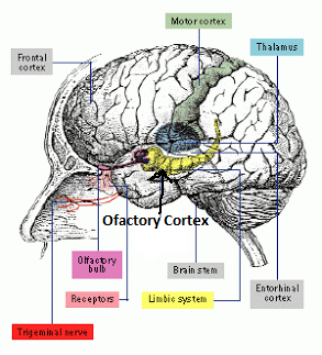
Textbook Module 7.3 Olfaction -Chemical Senses
STIMULUS
Olfaction is the response to chemicals that contact the membranes inside the nose. Olfaction is important in food selection, as most of what we call 'flavor" is a combination of the taste, texture and aroma of the food. The stimulus for olfaction are air-borne molecules that are ordorant chemicals
TRANSDUCTION
Olfactory cells line the nasal cavity. Each ell has cilia that extend from the cell body into the nasal passage. Olfactory receptors are located on the cilia. Ordorant chemicals activate synaptic transmission. Olfactory receptors have synapses with cells in the olfactory bulb.
While there are only 3 types of cone cells that produce color vision and 5 types of taste receptors that provide flavor, there are several hundred types of olfactory receptors that respond to different types of chemicals. Olfactory receptors are vulnerable to damage since they are exposed to the air. The lifetime for such cells is just around a month.

TRANSMISSION
When an olfactory receptor is stimulated the axon synapses in the olfactory bulb. Coding for the smell begins in the olfactory bulb. What does the book say about the changes in olfactory bulb cell firing based on whether the scent is different or more intense? KNOW FOR FINAL EXAM
Cells of the olfactory bulb axons synapse in the olfactory area of the cerebral cortex known as the olfactory cortex.

SENSATION PERCEPTION COGNITION
Olfaction also plays a role in social behavior. What are the scent-related chemicals involved in social behavior? Who detects orders more readily? Men or Women -- KNOW FOR FINAL EXAM
SYNESTHESIA
What is synesthesia? What sensory systems are identified in the textbook explaination of synesthesia? KNOW FOR FINAL EXAM
PUT IT ALL TOGETHER
For the final exam, you should know the answer to the questions in red above. Also you should be able to associate the following terms within the correct cell of the class schema.
air borne chemical ordorants
medial dorsal nucleus of the thalamus
nasal passages
Olfactory bulbs
olfacotry glomeruli
Olfactory protein molecule
olfactory receptor cells
orbitofrontal cortex
a rose
Olfaction is the response to chemicals that contact the membranes inside the nose. Olfaction is important in food selection, as most of what we call 'flavor" is a combination of the taste, texture and aroma of the food. The stimulus for olfaction are air-borne molecules that are ordorant chemicals
TRANSDUCTION
Olfactory cells line the nasal cavity. Each ell has cilia that extend from the cell body into the nasal passage. Olfactory receptors are located on the cilia. Ordorant chemicals activate synaptic transmission. Olfactory receptors have synapses with cells in the olfactory bulb.
While there are only 3 types of cone cells that produce color vision and 5 types of taste receptors that provide flavor, there are several hundred types of olfactory receptors that respond to different types of chemicals. Olfactory receptors are vulnerable to damage since they are exposed to the air. The lifetime for such cells is just around a month.

TRANSMISSION
When an olfactory receptor is stimulated the axon synapses in the olfactory bulb. Coding for the smell begins in the olfactory bulb. What does the book say about the changes in olfactory bulb cell firing based on whether the scent is different or more intense? KNOW FOR FINAL EXAM
Cells of the olfactory bulb axons synapse in the olfactory area of the cerebral cortex known as the olfactory cortex.
SENSATION PERCEPTION COGNITION
Olfaction also plays a role in social behavior. What are the scent-related chemicals involved in social behavior? Who detects orders more readily? Men or Women -- KNOW FOR FINAL EXAM
SYNESTHESIA
What is synesthesia? What sensory systems are identified in the textbook explaination of synesthesia? KNOW FOR FINAL EXAM
PUT IT ALL TOGETHER
For the final exam, you should know the answer to the questions in red above. Also you should be able to associate the following terms within the correct cell of the class schema.
air borne chemical ordorants
medial dorsal nucleus of the thalamus
nasal passages
Olfactory bulbs
olfacotry glomeruli
Olfactory protein molecule
olfactory receptor cells
orbitofrontal cortex
a rose
Sunday, March 22, 2015
Textbook Module 7.3 Gustation- Chemical Senses
CHEMICAL CODING
Most evolutionary scientists believe that the first sensory system of the earliest animal was a chemical sensitivity. Before the neocortex (Forebrain) evolved, there were areas of the midbrain and hindbrain devoted solely to sensing. Information on the Cranial Nerves are presented on page 84 (unit 5.7) of the coloring book.
What are the behavioral functions associated with the following cranial nerves (CN):
CN I.
CN II.;
CN V.
CN VIII
KNOW THE ANSWER TO THIS QUESTION FOR FINAL EXAM
STIMULUS
Organic substances containing chemicals that are perceived as sweet-sucrose, sour-HCL, bitter-quinine, salty-NaCl and umami or meaty-MSG. A combination of activity in five kinds of receptors (along with smell) give the perception of the taste of the food.
Taste buds are contained in papillae Taste receptors have excitable membranes and release neurotrnsmitters to excite other cells. Taste receptors are regenerated and replaced every 10-14 days, which is why food may taste more intense after an illness or after an extended fast. Taste receptors are located inside of tastebuds which are located in the papillae of the tongue.
TRANSDUCTION
When a substance tastes salty for instance, it means that a salitness receptor has detected the presence of sodium. Sodium ions cross into the membrane and produces an action potential. Sweetness, bitterness and umami receptors resemble the action of metabotropic receptors and therefore activate G-protein molecules and second messengers within the cell.

TRANSMISSION
Although each receptor detects one kind of taste, several receptors create a particular firing pattern that is perceived as a single taste experience. Information from the receptors in the anterior 2/3 of the tongue synpases with the Chorda Tympani nerve and information from the posterior tongue and the throat travel along branches of the Cranial Nervs IX and X. What is the name of Cranial Nerve X ?


Taste nerves project to the nucleus of the solitary track (NTS) in the medulla. From the NTS, the information branches out to the pons, the lateral thalamus, the amygdala and the ventral posterior thalamus (VPN), finally terminating into areas of the cerebral cortex. The somatosensory cortex is where touch or food texture is detected on the tongue and the insula or primary taste cortex is where taste is perceived.. Innervation is ipsilateral in this sensory modality.

SENSATION, PERCEPTION AND COGNITION
The sensation of taste can be affected by culture and familiarity but also by genes and hormones. Give examples from the textbook on how genetic differences affect taste. Also how do hormones affect taste preferences, according to the text? This is an extra credit opportunity and is due on April 2, 2015
PUT IT ALL TOGETHER
You should be able to associate the following terms within the correct cell of the class schema.
NaCl
Nucleus of the Solitary Tract
papillae
Primary gustatory cortex
saliva
taste buds
taste receptors
teeth
tongue
unami
Vagus Nerve
Ventral Posterior nucleus
Most evolutionary scientists believe that the first sensory system of the earliest animal was a chemical sensitivity. Before the neocortex (Forebrain) evolved, there were areas of the midbrain and hindbrain devoted solely to sensing. Information on the Cranial Nerves are presented on page 84 (unit 5.7) of the coloring book.
What are the behavioral functions associated with the following cranial nerves (CN):
CN I.
CN II.;
CN V.
CN VIII
KNOW THE ANSWER TO THIS QUESTION FOR FINAL EXAM
STIMULUS
Organic substances containing chemicals that are perceived as sweet-sucrose, sour-HCL, bitter-quinine, salty-NaCl and umami or meaty-MSG. A combination of activity in five kinds of receptors (along with smell) give the perception of the taste of the food.
Taste buds are contained in papillae Taste receptors have excitable membranes and release neurotrnsmitters to excite other cells. Taste receptors are regenerated and replaced every 10-14 days, which is why food may taste more intense after an illness or after an extended fast. Taste receptors are located inside of tastebuds which are located in the papillae of the tongue.
TRANSDUCTION
When a substance tastes salty for instance, it means that a salitness receptor has detected the presence of sodium. Sodium ions cross into the membrane and produces an action potential. Sweetness, bitterness and umami receptors resemble the action of metabotropic receptors and therefore activate G-protein molecules and second messengers within the cell.

TRANSMISSION
Although each receptor detects one kind of taste, several receptors create a particular firing pattern that is perceived as a single taste experience. Information from the receptors in the anterior 2/3 of the tongue synpases with the Chorda Tympani nerve and information from the posterior tongue and the throat travel along branches of the Cranial Nervs IX and X. What is the name of Cranial Nerve X ?

Taste nerves project to the nucleus of the solitary track (NTS) in the medulla. From the NTS, the information branches out to the pons, the lateral thalamus, the amygdala and the ventral posterior thalamus (VPN), finally terminating into areas of the cerebral cortex. The somatosensory cortex is where touch or food texture is detected on the tongue and the insula or primary taste cortex is where taste is perceived.. Innervation is ipsilateral in this sensory modality.

SENSATION, PERCEPTION AND COGNITION
The sensation of taste can be affected by culture and familiarity but also by genes and hormones. Give examples from the textbook on how genetic differences affect taste. Also how do hormones affect taste preferences, according to the text? This is an extra credit opportunity and is due on April 2, 2015
PUT IT ALL TOGETHER
You should be able to associate the following terms within the correct cell of the class schema.
NaCl
Nucleus of the Solitary Tract
papillae
Primary gustatory cortex
saliva
taste buds
taste receptors
teeth
tongue
unami
Vagus Nerve
Ventral Posterior nucleus
Monday, January 12, 2015
Textbook Module 1.1 Biological Approach to Behavior
Module 1.1 Biological Approach to Behavior- January 13, 2015
Lecture Notes
I. INTRODUCTION Textbook pg 2"Why is there something rather than nothing?"
Science studies the universe on many different levels of analyses. See powers of ten On the EXPANDING view of Powers of Ten we get a picture of our place in the universe.
What does the COLLAPSING view indicate to you?
The film simply allow us to see what is possible to observe and speculate the universe as far away and as deep down as humans technology can take us. The technology is just one aspect of human curiosity that allows the asking of questions. David Chalmers, a philosopher says that the "hard problem" is ...
"Why is there such a thing as consciousness?"
A. Biological Psychology- the study of the physiological, evolutionary and developmental mechanism of behavior and experience and is similar to: Biopscychology, psychobiology, physiological psychology and behavioral neuroscience.
CAN YOU ANSWER THIS QUESTION-
? What is the difference between biological psychology and behavioral neuroscience.?
B. Two views of the human brain (Figure 1.2 pg 4)
outer view
| DORSAL | VENTRAL |
| Frontal Lobe | Frontal Lobe |
| Pre central Gyrus | Temporal lobe |
| Central Sulcus | Longitudinal fissure |
| Post central Gyrus | Olfactory Bulb |
| Parietal Lobe | Optic nerve |
| Occipital Lobe | Medulla, Cerebellum and Spinal cord |
CAN YOU IDENTIFY MOST OF THESE AREAS FROM THE COLORING BOOK?- 7.1,7.2 AND 7.3
inner view
Figure 1.3 Neurons magnified
CAN YOU IDENTIFY THE TRANSMITTING AND RECEIVING ENDS OF THE NEURON?
COLORING BOOK -3.1
II. Biological Explanations of Behavior
The four categories of behavior- according to the biological explanation of behavior.
A. Physiological explanation
B. Ontogenetic explanation
C. Evolutionary explanation
D. Functional explanation
CLOSE BOOK-
Explain the birdsong in terms of the four categories.
III. Career Opportunities
Q. How many different fields of Psychology are there?
A. Research, Practioner, Medical Allied Medical
Q. What particular specialization seems most interesting to you? Why?
ADDITIONAL FACTS FROM 12TH EDITION-
There are three main points that the author wants you to remember from the book.
#1 "Perception occurs in your brain."
#2 "Mental activity and certain types of brain activity are, "So far as we can tell, inseparable." This view is known as monism. (This is the view of the author but not all scientists agree. See here for an alternative view to monism from Biologist Richard Sheldrake.
#3 Don't confuse an example with an explanation. Examples of scientific findings can convey many different things and never will a single study provide all the answers.
Saturday, January 10, 2015
Module 2.1: Cellualr Morphology-Anatomy of Neurons and Glia Cells
Anatomy of Neurons and Glia- MODULES 1.2 & 2.1
A The histology of Santiago Ramon y Cajal
Cajal was both a physician and an artist. He used his skills to draw images that he saw under the microscope with the help of Golgi staining. Both he and Golgi received the 1906 Nobel Prize. Go here to view some of his drawings.
Q. What is "histology?"
B. Learn the parts of the neuron.
Using "Google Image" find a few pictures of the Neuron. These pictures will assist you as you color the pages of Coloring book chapter 3.
Q. Can you match the pictures, terms and definitions for the following from the coloring book:
ASSIGNMENT #1
3.1: cell body (soma), dendrites, axon, cytoplasm, interstitial fluid, cell membrane (lipid bilayer) terminal buttons (boutons) axon branches (axon terminal)
3.2: nucleus, neurofilaments microtubules
3.3: neurotransmitter molecules; synaptic vesicles
3.4 synapase, receptive area (dendritic arborization), axon hillock, axon shaft
ASSIGNMENT #2
3.5: pre-synaptic membrane, post-synaptic memberane, receptors, neurtransmitter molecule, synaptic vesicle, omega complex
3.6: schwann cells, nodes of Ranvier, Oligodendrocyte myelin sheath
ASSIGNMENT #3
3.7 ion channels, ionotropic receptor
III. The Blood Brain Barrier
Q. How does the brain blood barrier work?
IV. Nourishment in Vertebrate Neurons
Q. What is the importance of glucose to the brain?
Q. Define a vertebrate neuron. (Hint: go to CB unit 1.1)
Wednesday, January 7, 2015
Textbook Module 4.1 Anatomy of the Nervous System- February 6, 2014
Textbook Module 4.1
Please begin coloring the pages associated with Module 4.1. The assignments can be found in the coloring book assignment link at the top of the page.
Please begin coloring the pages associated with Module 4.1. The assignments can be found in the coloring book assignment link at the top of the page.
Monday, January 5, 2015
Practice Homework Gross Anatomy
Answer these questions after you have colored the following coloring book units.
1. What type of section is illustrated in CBUnit 10.5B?2. What type of section is illustrated in CB Unit 11.6B
3. What type of section is illustrated in CB Unit
12.5?
4. Name the
most posterior lobe of the brain.
5.
Name the most lateral lobe of the brain.
6.
Name the most anterior lobe of the brain.
7. Which lobe has the post-central gyrus and central fissure as a
boundary?
8. Which lobe has the superior temporal gyrus and lateral fissue as a boundary?
9.
Name the fissure that separates the brain
hemispheres.
10. Distinguish by location the differences between the superior temporal gyrus and the superior frontal gyrus. (ie which brain lobe contains each gyri).
10. Distinguish by location the differences between the superior temporal gyrus and the superior frontal gyrus. (ie which brain lobe contains each gyri).
Textbook Module 3.1 Synpatic Transmission
Lecture material
Properties of the synapse pg 52-58

Synaptic transmission (neuron story) explains in a very simplified form how neurons communicate with each other. In the illustration imagine that the stimulus is a fly landing on your leg and the response is moving the leg. Identify Cell A, Cell B and Cell C.
Reproduce the A-B-C neural network on page 9 (Unit 1.4) of the coloring book.
Color Coloring Book Units 1.3, 1.4, 1.5 and 1.6 ( pgs 8-15)
Review exercise 1.1, 1.2 and 1.3 on page 16-17
What is the difference between temporal and spatial summation.
Relationship Among EPSP, IPSP and action Potentials Pgs 56-57
Explain the role of the interneuron in an inhibitory synapse- use Figure 3.7 pg 56.
Create a wiring diagram that shows inhibition and excitation... use figure 3.9; 3.10 and 3.11 as examples but create your own using any configuration.
Subscribe to:
Comments (Atom)